
#EEG ELECTRODE PLACEMENT FULL#
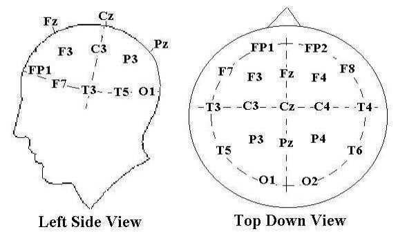
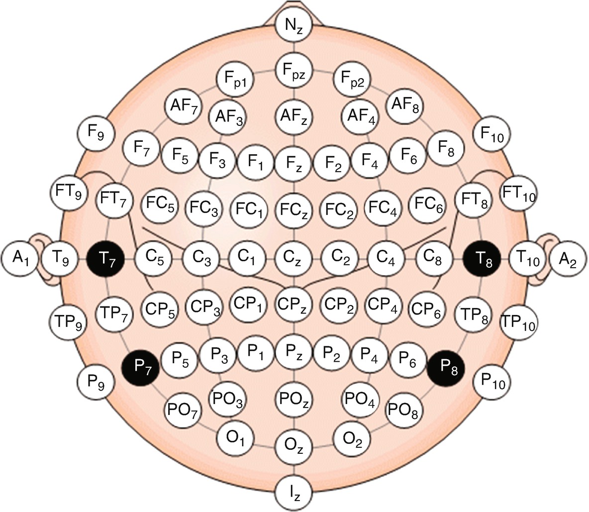
There is a discharge of -50mV, but depending on its dipole and location, not every electrode will see a full -50mV. To better understand how bipolar montages lead to EEG tracings, lets take a look at the example below. For example, if Fp2 has a voltage of -50 and F8 has a voltage of -20, the Fp2-F8 tracing would show -50 - (-20) = -30mV. Because of this, in bipolar if the first electrode in the tracing line is more positive/higher than the second, you get a positive, downward deflection if the second electrode is more positive/higher, you get a negative, upward deflection. In each chain, an electrode's voltage is compared to that of the electrode behind it, so each tracing line is a pair of electrodes in which the voltage of the second electrode is subtracted from the voltage of the first. From there, you divide the head into consecutive increments of 10% and 20% to find the placement of the electrodes, as shown in the illustration below (which is not, of note, anatomically correct!). The first step in the 10-20 system setup is finding the nasion (the top of the nose bride, between the eyes) and the inion (the small bump in the middle of the back of the head). For the midline/central electrodes, instead of a number their letters are clarified with a " z." The number corresponds to the side of the brain- odds on the left, evens on the right-and the particular area of each region. The letter corresponds to its region of the brain: F for frontal region, T for temporal, P for parietal, and O for occipital (the only exception to this is that F7 and F8, while seemingly frontal, are in fact actually over the anterior temporal region). In the 10-20 system, each electrode is identified with a letter and a number. This system is so named because it splits the skull into increments of 10% or 20% to place the electrodes, ensuring that each electrode is relatively positioned to all the others and making it possible for every EEG study to be consistent despite peoples' many different head shapes and sizes. The first step to any EEG study is the placement of the electrodes, and this is most commonly done via the international, standardized 10-20 System.

Now that you have a grasp of the pertinent neurophysiology that gives rise to the EEG signal, let's finally dive into the EEG itself.


 0 kommentar(er)
